Shape Analysis Group

Nuclear Patterning of Developing Cells in Murine Ventricular Heart Walls
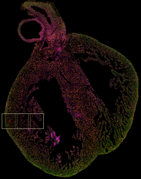
Figure: A model for orthogonal myofiber systems in heart ventricular walls.
In embryonic stages of development, the mammalian heart is characterized by a rapid division of cells, and their organization into a functional beating organ. Although cardiomyocyte nuclei are known to be elongated and oriented along their length in the adult heart, how nuclear shape, orientation, and arrangement evolve during development is not well understood. To tackle this fundamental problem we examine the evolution of nuclear geometry in the mouse heart wall over four post-septation embryonic days, and in the newborn pup, using confocal microscopy and quantitative modeling. Over this period there are notable changes in nuclear count, density, spatial layout, and organization. For example, we observe a steady increase in nuclear elongation, but a decrease in their volume. Whereas nuclei are more densely packed towards the epicardium just after septation, in time they become more uniformly distributed throughout the heart wall. We hypothesize that such structural arrangements in the developing myocardium could have an important bearing on healthy heart function.